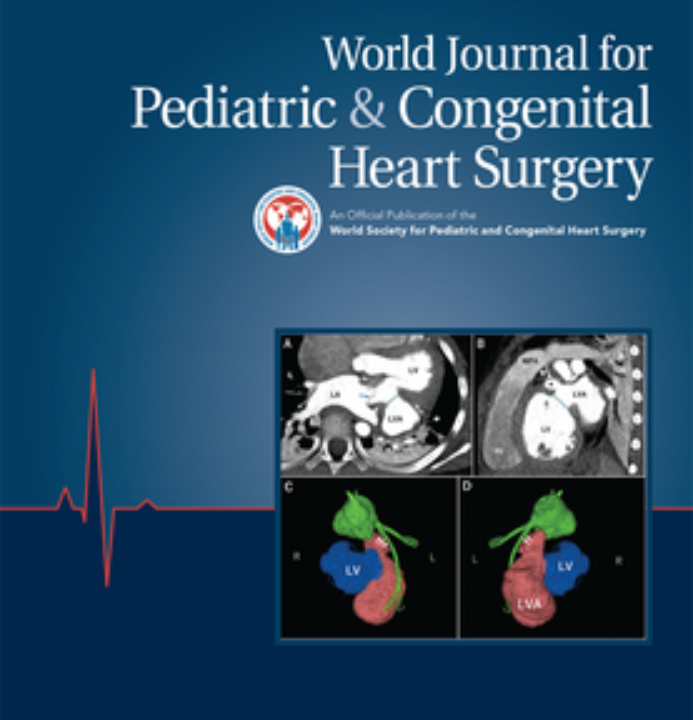
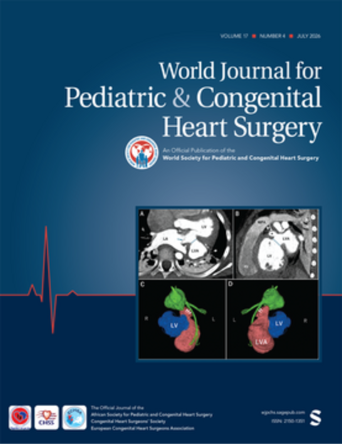
Infracardiac Total Anomalous Pulmonary Venous Connection with Meandering Intrapulmonary Vertical Vein Masquerading as a Scimitar Vein
Published: February 21, 2025.
Online first
Contributors
Damandeep Singh; Niraj Nirmal Pandey; Aprateem Mukherjee; Ankur Handa; Lamk Kadiyani; Vatsal Vora; Pradeep Ramakrishnan; Sanjeev Kumar
World Journal for Pediatric and Congenital Heart Surgery, Ahead of Print.
Read this article | Join for full access
Conduit Size, Branch Pulmonary Artery Size, and Reoperation in Patients With Truncus Arteriosus
Published: February 21, 2025.
Online first
Contributors
Christopher W. Mastropietro; Andrew Rodenbarger; Jeremy L. Herrmann
World Journal for Pediatric and Congenital Heart Surgery, Ahead of Print.
BackgroundOptimal right ventricle-to-pulmonary artery (RV-PA) conduit size for patients with truncus arteriosus is controversial. We aimed to determine the relationship between branch PA size and the need for conduit reoperation following repair of truncus arteriosus.MethodsWe performed a single-center chart review of patients who underwent truncus arteriosus repair from January 2009 to December 2023. Branch PAs were measured in systole, at the narrowest point if focal stenosis was present. For branch PA diameter analyses, the smaller diameter PA was used. Univariate Cox proportional hazards regression analysis was performed to determine hazard ratios (HRs) with 95% confidence intervals (CIs) for echocardiographic measures and conduit reoperation.ResultsWe included 33 patients. Median age at surgery was 23 days (range: 3–34). Thirty-two patients received a bovine jugular vein graft, one patient received an aortic homograft. Mean RV-PA conduit Z-score was 2.7 ± 0.5 and mean preoperative conduit-to-PA ratio 2.6 ± 0.6. Postoperative diameter of at least one branch PA was decreased in 31 patients (93.9%); mean change was −19% ± 17%. Mean postoperative conduit-to-PA ratio was 3.3 ± 0.9. Conduit reoperation occurred in 19 patients (58%); median time to reoperation was 1.6 years (range: 0.4-10.4). Conduit reoperation was not associated with conduit diameter or Z-score. Conduit reoperation was significantly associated with truncus type A2 or A3 PA anatomy (HR: 3.53; 95%CI: 1.14-10.94) and conduit-to-PA ratio ≥ 4 (HR: 4.94; 95%CI: 1.63-14.97).ConclusionIn a single-center cohort of children who underwent repair of truncus arteriosus, RV-PA conduit diameter was not associated with increased conduit longevity. Rather, larger postoperative RV-PA conduit to branch PA diameter ratio was significantly associated with greater hazard for conduit reoperation.
Read this article | Join for full access
Response to Reader Comment on: A Low-Cost Workflow to Generate Virtual and Physical Three-Dimensional Models of Cardiac Structures. If You Can Make it Here, You Can Make it Anywhere
Published: February 21, 2025.
Online first
Contributors
Alexander Schneller; Philippe Grieshaber
World Journal for Pediatric and Congenital Heart Surgery, Ahead of Print.
Read this article | Join for full access
Predictors of Success Following Valve-Sparing Repair of Tetralogy of Fallot
Published: February 21, 2025.
Online first
Contributors
Paighton C. Miller; Michael R. Chomat; Fei Wan; Jacob R. Miller; Dilip Nath; Pirooz Eghtesady
World Journal for Pediatric and Congenital Heart Surgery, Ahead of Print.
Objective: We aim to report predictors of success following valve-sparing repair (VSR) for Tetralogy of Fallot (TOF). Methods: We performed a single-institution retrospective review of 70 patients who underwent VSR for TOF from 2007 to 2021. Risk factors for moderate to severe pulmonary insufficiency (PI) and surgical or catheter intervention for right ventricular outflow tract (RVOT) obstruction were analyzed. Results: During a median follow-up time of 6 years (range 1 month to 17 years), 5/70 (7%) patients required surgical or catheter intervention for isolated RVOT obstruction, 8/70 (11%) had moderate or severe PI, and 3/70 (4%) had a combined outcome of RVOT obstruction and PI. Patients who required reintervention had smaller pulmonary valve (PV) z-score (−2.8 vs −1.7, P < .01), were more likely to have isolated infundibular patching (75% vs 31%, P = .02), and had smaller PV z-score at the end of the procedure (−1.4 vs −1.0, P = .03). Patients with significant PI were more likely to have intraoperative valvotomy via Hegar dilation (36% vs 13%, P = .04). The strongest independent predictors of RVOT obstruction and/or PI were preoperative cyanotic episodes (odds ratio 6.0, 95%CI: 1.6-22, P = .01) and valvotomy via Hegar dilation (odds ratio 4.6, 95%CI: 1.0-21, P = .04). Conclusions: Valve-sparing repair of TOF is less likely to be successful if reliant on isolated infundibular patching or not achieving at least a z-score of −1 at the PV at the completion of the procedure. Repairs using blind dilation destabilize the valve and lead to long-term valve incompetence.
Read this article | Join for full access
Rehabilitation Strategy Should Not Be a Pretext for Suboptimal Repair for Pulmonary Atresia, Ventricular Septal Defect, and Major Aortopulmonary Collateral Arteries
Published: February 21, 2025.
Online first
Contributors
Manan H. Desai; Aybala Tongut; Yves d’Udekem
World Journal for Pediatric and Congenital Heart Surgery, Ahead of Print.
Pulmonary atresia, ventricular septal defect, and major aortopulmonary collaterals are a challenging congenital anomaly to manage surgically with different centers and strategies producing wide-ranging outcomes. Over the past few decades despite diverging treatment pathways, there is an emerging consensus of how these patients “should” be treated. Quite often a combination of rehabilitation strategy and a unifocalization approach has to be tailored to each patient to address the anatomic and physiological variations that characterize this congenital heart defect. Irrespective of the surgical approach, the goal should be to have a complete repair with acceptable right heart pressure ensuring survival and a good quality of life.
Read this article | Join for full access
Tetralogy of Fallot With Pulmonary Atresia and Major Aortopulmonary Collateral Arteries: Diagnostic Modalities—the Role of Computed Tomography, Cardiac Magnetic Resonance Imaging, and Three-Dimensional Modeling
Published: February 21, 2025.
Online first
Contributors
Reena M. Ghosh; Mara Thompson
World Journal for Pediatric and Congenital Heart Surgery, Ahead of Print.
Cross-sectional imaging modalities facilitate the noninvasive acquisition of anatomic and hemodynamic data in patients with congenital heart disease. This is critical in patients with tetralogy of Fallot with pulmonary atresia and major aortopulmonary collateral arteries (TOF PA MAPCAs), who typically undergo multiple surgeries and interventional catheterizations over the course of their lifetime. Advances in cardiac computed tomography (CT) and cardiac magnetic resonance imaging (CMR) have enabled increased spatial resolution imaging and decreased scan times. High spatial resolution datasets provide a comprehensive evaluation of MAPCA anatomy including aortic origin location, lung lobes supplied, and the presence of stenoses. The physiologic data provided by CMR offers a noninvasive assessment of both anatomy and pulmonary blood flow, which can be utilized in decision-making and patient risk stratification. Furthermore, the three-dimensional (3D) datasets acquired by either imaging modality can be used to derive a 3D representation of patient-specific anatomy, visualized in a variety of formats including on-screen volume rendering, 3D prints, digital 3D models, and virtual reality. This article reviews the utility and added value of cardiac CT, CMR, and 3D modeling in the diagnosis and management of patients with TOF/PA/MAPCAs.
Read this article | Join for full access
Long-Term Outcomes After Pulmonary Valve Repair for Regurgitation Secondary to Prior Intervention
Published: February 21, 2025.
Online first
Contributors
Daniel Kyrillos Ragheb; Yulin Zhang; Ayush Jaggi; Shiraz A. Maskatia; Gregory T. Adamson; George K. Lui; Elisabeth Martin; Michael Ma; Frank L. Hanley; Doff B. McElhinney
World Journal for Pediatric and Congenital Heart Surgery, Ahead of Print.
PurposePulmonary valve (PV) regurgitation (PR) secondary to prior repair of congenital heart disease commonly necessitates intervention, typically with PV replacement (PVR). However, prosthetic valves are susceptible to degeneration and ultimately require reintervention. Pulmonary valve repair (PVr) can correct PR while retaining native tissue, but long-term durability is unknown.MethodsAll patients who underwent PVr from 2010 to 2018 for PR resulting from prior PV intervention were included. A control cohort included patients who underwent PVR during the same period for the same indications. Time-related outcomes including freedom from right ventricular outflow tract reintervention, moderate or greater PR, and a maximum Doppler gradient ≥36 mm Hg were compared. Approved as IRB-65340.ResultsThe study included 33 and 151 patients who underwent PVr and PVR, respectively, 72% (132/184) with tetralogy of Fallot. Patients were followed for a median of 9.0 years (6.4-11.5) and 7.7 years (5.4-9.9), respectively (P = .041). Estimated freedom from reintervention 5 and 10 years after discharge was 97% (80-100) and 89% (69-96) after PVr and 96% (92-99) and 79% (67-87) after PVR. On Cox regression analysis adjusted for age or weight at the time of surgery, and on multivariable Cox regression, PVr was associated with significantly longer freedom from reintervention and valve dysfunction than PVR.ConclusionsPulmonary valve repair was associated with longer freedom from valve dysfunction and reintervention than PVR, particularly in pediatric patients. Lifetime management should be considered at original repair, with an effort to maintain native tissue for potential future PVr.
Read this article | Join for full access
Native Tricuspid Valve Endocarditis Delayed Glenn Procedure: A Case Report
Published: February 20, 2025.
Online first
Contributors
Madison B. Argo; Joshua L. Hermsen; Luke Lamers; Mike Wilhelm; Charles Bergstrom; Petros V. Anagnostopoulos
World Journal for Pediatric and Congenital Heart Surgery, Ahead of Print.
Infective endocarditis in infants with congenital heart disease is exceedingly rare and thus, treatment guidelines and expected outcomes are understudied. We present a 3-month-old girl diagnosed with native tricuspid valve endocarditis at the time of her pre-Glenn cardiac catheterization. She had resultant tricuspid valve and right ventricular dysfunction. After initiating intravenous antibiotics, she underwent an operation including a bidirectional Glenn, Sano shunt extirpation, tricuspid valve repair, and central pulmonary artery stent insertion. At 9 months postoperatively, the patient continues to do well with good right ventricular and tricuspid valve function. Surgical treatment of infective endocarditis may be performed during a Glenn operation with satisfactory short-term results.
Read this article | Join for full access
Routine Use of an On-Table Extubation Protocol in Pediatric Cardiac Surgery—Our Experience With Life in the Fast Lane
Published: February 13, 2025.
Online first
Contributors
Jothinath Kaushik; Raju Vijayakumar; Pavithra Ramanath; Murugesan Karthik Babu; Srinivasan Naveen; Janarthanan Maniyarasu; Michael E. Nemergut; Joseph Dearani
World Journal for Pediatric and Congenital Heart Surgery, Ahead of Print.
BackgroundWe undertook this study to evaluate the efficacy of an on-table extubation protocol and to assess the magnitude of benefits when implemented as a routine practice in a developing country.MethodsThis prospective observational study at a single tertiary care referral hospital was designed to determine the efficacy of an on-table extubation protocol when applied to children undergoing cardiac surgery in the developing world. The study included 226 patients who were 1 month to 18 years of age undergoing cardiac surgery (including grown-up congenital heart disease [GUCHD] patients). Patients with RACHS score ≥ 4, neonates, preoperatively ventilated children, and emergency surgeries were excluded from the study. All pediatric elective cardiac surgical patients belonging to RACHS 1, 2, and 3 categories were considered as potential candidates for on-table extubation. Trial registration: Clinical Trials Registry of India (CTRI/2020/07/026567).ResultsAmong the 226 children who underwent elective cardiac surgeries, we were able to extubate 142 patients (62.83%) in the operating room. This included 46.6% (54/116) infants, 80.8% (38/47) children less than 5 years of age, 79.3% (46/58) children between 5 years to 18 years age, and 80% (4/5) GUCHD. The duration of intensive care unit (ICU) stay, hospital stay, and hospital cost were significantly less in the on-table extubation group (23 [20, 26] hours; 102 [97, 125] hours; INR 2,09,011 [181032, 244298]) as compared with those patients extubated in the ICU within 6 hours (28 [22, 46] hours; 122 [100, 168] hours; INR 2,25,430 [162203, 273831]) and beyond 6 hours (71 [45, 121] hours; 184 [127, 243] hours; INR 2,53,541 [226838, 306871]).ConclusionsThis protocol shows a significant reduction in ICU stay, hospital stay, and total hospital cost when compared with either extubation within 6 h in the ICU or delayed extubation (beyond 6 h) in patients undergoing pediatric cardiac surgery.
